Taxonomic Tools: Scanning Electron Microscope (SEM)
Traditional optical microscopes direct beams of light through a series of lenses to magnify an object, but they are limited to about 1,500X magnification. In taxonomy, the science of identifying and classifying organisms, key morphological traits are sometimes too small to be seen using these instruments. Scanning Electron Microscope (SEM) is a powerful imaging tool that uses electrons instead of light to generate highly detailed images of microscopic structures, beyond what a light microscope can achieve. SEM is especially valuable to observe, measure, and describe some incredibly small and intricately ornamented structures of invertebrates that are crucial for identifying and classifying species.
The Scientific Imaging facilities at the National Museum of Natural History house two SEM microscopes. In the context of the MDBC project scientists have been using these microscopes to study calcareous sclerites, which are mineralized structures found in certain animals like sponges, corals, and mollusks. These structures are essential for identifying species of bamboo corals, black corals, sponges, and solenogasters (shown below).
SEM
Scales of a new species of Solenogaster - a worm-like shell less mollusk - Dondersiidae sp. nov. Sclerites are important to distinguish between species of this family. While they are impossible to see with the naked eye, SEM allows experts like postdoc Carmen Cobo to see the shape and intricate arrangement of the scales.
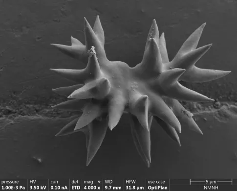
Sponge’s spicules - Acanthochela of a new species. This tiny structure measures just 15 micrometers in length and exhibits intricate ornamentation. To put that in perspective, a human hair is about 70 micrometers thick, so this sponge spicule is almost five times smaller than a strand hair! Such minute details are impossible to observe with standard optical microscopy but are clearly revealed under the powerful magnification of a SEM. By using SEM images, we can measure, describe, and compare spicules in detail, which is essential for identifying and classifying sponge species.
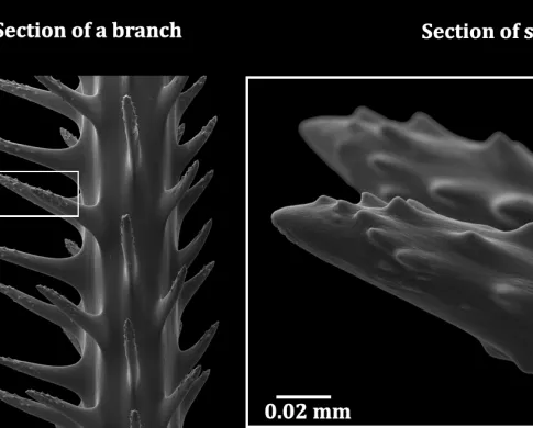
Branches of Aphanipathes puertoricoensis (Horowitz & Quattrini, 2023) (left) with a highlighted section showing the branch skeleton (middle) and a zoomed-in view of skeletal spines and tubercles (right). These small surface features aid in identifying and describing black coral species and can only be seen using scanning electron microscopy.
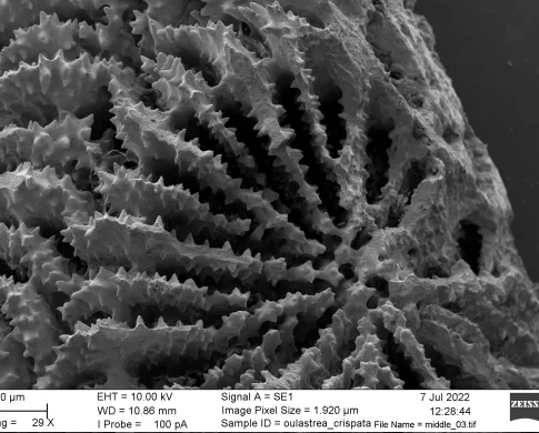
Septa details of a polyp of the species Oulastrea crispata. The arrangement of the septa and their ornaments can aid in understanding species relationships with closely related taxa.
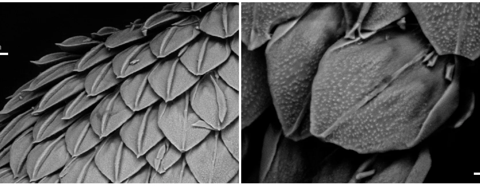
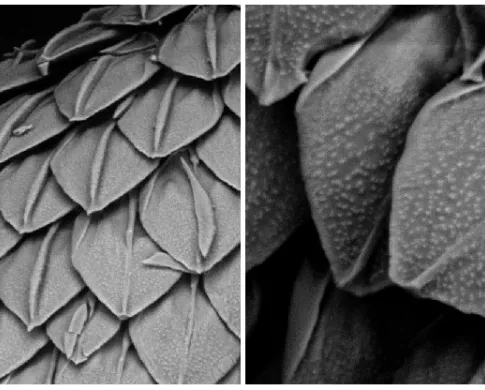
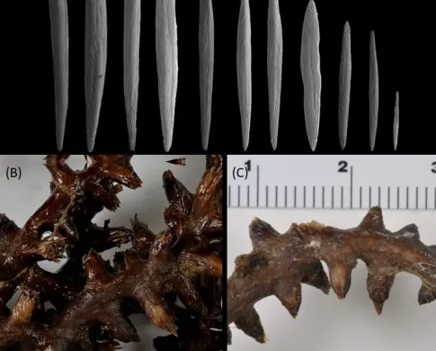
Explorisis katharina (Anthozoa, Octocorallia) (A) Sclerites from the polyps, (B-C) close up of polyps from Explorisis katharina. Scale bar is 1mm. The shape and composition of the sclerites in different parts of the coral can help us distinguish both the species and genus the corals belong to.